What Causes Vitreomacular Traction?
Vitreomacular traction is typically caused by the vitreous not detaching completely from the retina as you age (PVD). Some factors may increase your risk of developing vitreomacular traction, including:
- Age-related macular degeneration (AMD): AMD is a condition common with aging in which the macula begins to deteriorate.
- Diabetic retinopathy and diabetic macular edema: Diabetic retinopathy is when diabetes causes damage to the blood vessels in the retina. Diabetic retinopathy can lead to proliferative diabetic retinopathy (PDR) — when abnormal blood vessels grow on the retina — and diabetic macular edema, a condition where fluid builds up in the macula. These conditions also increase your risk of vitreomacular traction.
- Extreme nearsightedness
- Retinal vein occlusion: In a retinal vein occlusion, one of the veins leading out of the eye becomes blocked
What Are the Symptoms of Vitreomacular Traction?
Vitreomacular traction often leads to changes in vision. These may include:
- Decrease in vision sharpness
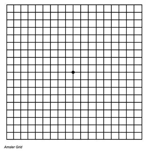
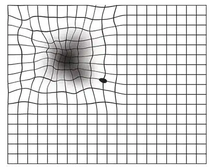
- Distortions in vision that make straight lines look wavy (metamorphosis)
- Objects looking smaller than their actual size (micropsia)
- Seeing flashes of light in the eye (photopsia)
These symptoms may come on slowly. They also mimic many other eye conditions, so it’s important to see a doctor if you experience these symptoms.
How Is Vitreomacular Traction Diagnosed?
Your ophthalmologist will need to look inside your eye in order to diagnose vitreomacular traction. There are a few different imaging tests they can use to do this, including:
- Dynamic B-scan ultrasound: This is a type of ultrasound, which is a test that uses sound waves to create an image of the inside of the body. Dynamic B-scan ultrasound can show the doctor what the relationship between the vitreous and retina looks like.
- Fluorescein angiography: In fluorescein angiography, your doctor injects yellow dye into your arm. This dye travels to the blood vessels of your eye, and your doctor uses a special camera to take pictures of the eye. The dye shows up brightly on the photos, helping your doctor see where the issues may be.
- Optical Coherence Tomography: Optical coherence tomography is the test most commonly used to diagnose vitreomacular traction. This test uses light waves to create cross-sectional images of the layers in the retina.
How Is Vitreomacular Traction Treated?
There are generally four treatment options used to treat vitreomacular traction: monitoring, medication, pneumatic vitreolysis, and surgery. Your doctor will recommend a treatment option based on how severe your vitreomacular traction is.
Vitrectomy Surgery
In severe cases, your ophthalmologist may decide to perform a type of surgery called a vitrectomy.
A vitrectomy must be done in a surgery center. Your doctor will make a small cut into your eye and use a microscope to see inside your eye. They then use very small tools to sever the connection between the vitreous and retina and repair any damage to the retina.
Pneumatic Vitreolysis
Pneumatic vitreolysis is a procedure where your doctor injects a small gas bubble into your eye. The goal is to get the bubble to break the bond between the vitreous and the macula. To make that work, you will need to look down several times an hour for a few days to get the bubble into the right position.
Sometimes, your doctor will use pneumatic vitreolysis in combination with medication to encourage the vitreous to fully separate from the macula.
Medication
A medication called ocriplasmin is proven to be a good option for people with vitreomacular traction. It works by dissolving the fibers that keep the vitreous stuck to the macula. Ocriplasmin is given by a single injection into the center of the eye.